ARTICLE AD BOX
 Image source, HELMHOTZ MUNICH
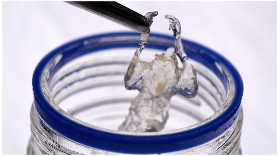
Everything inside the mouse – its nerves, tissues and organs – are made invisible by a chemical process
By Pallab Ghosh
Science correspondent
A new scanning method involving a see-through mouse could improve how cancer drugs are tested, by picking up tumours previously too small to detect.
Prof Ali Ertürk of the Helmholtz Munich research centre worked out how to make a dead mouse transparent in 2018.
His team have now used chemicals to highlight specific tissues so that they can be scanned in unprecedented detail.
Drugs are often tested first on mice. Scientists say the new scanning method could revolutionise medical research.
Cancer Research UK said the new scanning technique had "a wealth of potential".
Watch: Scanning a transparent mouse to reveal the body in unprecedented detail
The researchers say the method reveals far greater detail than existing scanning techniques. In one of the first applications the team has detected cancerous tumours in the first stages of formation.
Prof Ertürk says this is important because cancer drugs have to be shown to eliminate tumours in mice before being tested on humans.
"MRI and PET scans would show you only big tumours. Ours show tumours at the single cell, which they absolutely can't".
"Current drugs extend life by a few years and then the cancer comes back. This is because the development process never included eliminating those tiny tumours, which were never visible."
Normally lab mice are given cancer and scanned with conventional scans to see how the tumour has progressed. They are then treated with the cancer drug being tested and then scanned again to see what if any difference the treatment has had.
Prof Ertürk's scanning method can only be used on dead mice, to give a picture of how much cancer has progressed, or potentially, whether a treatment has worked. He made mice transparent after they were given cancer and then scanned them using his new technique. Only a few mice would need to be made transparent to test the effectiveness of the drug.
Image source, ALI ERTURK/NATURE
Image caption,Scanning (below in blue) shows cancer tumours as pink and white dots. A conventional scan (in white) shows only the largest concentrations of tumours.
Dr Rupal Mistry, research information manager at Cancer Research UK, said:
"This exciting and unique scanning technique has a wealth of potential for building our knowledge of how our bodies work and what goes wrong in diseases like cancer.
"While researchers will only be able to use the technique to examine the bodies of deceased mice, it could tell us a lot about how cancer develops at the early stages of the disease. Being able to visualise tumours in the context of the entire body will also give researchers a greater understanding of the impact of different drugs and treatment.
"Advances in technology like this are essential to driving progress and will hopefully lead to new ways to detect, treat and prevent cancer."
The cancer application, published in the journal Nature Biotechnology, is just one of hundreds if not thousands to which the new scanning technique can be used to improve medical studies. It can enable researchers to see things they have never seen before.
Mouse studies are often the starting point for learning about processes in the human body. But the new technique can be used on any animals. It could also be used to make human tissues and organs transparent, though it is unlikely to be used to make an entire human body transparent in the near future because there would be no medical advances that could be made from it at this stage.
Creation of the transparent mouse involves removing all the fats and pigment from its corpse, using a chemical process. It ends up looking like a clear plastic toy, which is ever so slightly bendy. Its organs and nerves are all still inside it - but near invisible.
While Prof Ertürk's developed the process to make a mouse transparent five years ago, the scanning technique makes the most of it.
He has found a way of adding other chemicals known as antibodies to highlight the parts of the mouse he is interested in studying under a microscope. Different antibodies stick to different types of tissue and so highlight whatever the researchers are interested in looking at.
As well as highlighting cancerous areas, Prof Ertürk's team has produced a suite of videos which enable researchers to fly through the mouse's nervous system, gut or lymph system.
The scans have several advantages over what is available now.
First, the researchers can study diseases in the context of the entire body, which gives them a much greater understanding of the impact of different drugs and treatments.
The 3D images are also stored online, so researchers studying different parts of the animal or wanting to do the same experiment can draw from a library, rather than having to use another mouse. Prof Ertürk believes that the technique could reduce lab animal use tenfold.
Image source, HELMHOTZ MUNICH
Image caption,Prof Ali Ertürk about to dip a transparent mouse into chemicals that highlight specific tissues
Dr Nana-Jane Chipampe, of the Wellcome Sanger Institute in Cambridge, is excited at the prospect of using the new scanning technique to study how cells develop in the human body. Currently she has to slice up tissues into very thin sections to study them under a microscope. Soon she will be able to see details in 3D.
"I can't wait to get my hands on it!" she told me enthusiastically.
"It has the potential to identify new tissues, cells and diseases which will really help us understand the development of diseases."
Her team leader, Prof Muzlifah Haniffa, is producing an online map or atlas of every cell in the human body. She says the new scanning technique will be useful for all kinds of medical research.
"Without a doubt, it will accelerate the pace of medical research," she said. "Combining these cutting-edge technologies and building the human cell atlas will no doubt completely revolutionise medicine."

 1 year ago
46


 English (US) ·